2011 article
Source michaeldmann.net
Photoreception is a particularly important sense for most primates, including man, but it is not unique to primates or even mammals. Even mollusks have photoreceptors, but one may question whether they possess vision in the same sense as we have it. Most objects reflect light, and because light travels at high speed, it is possible to nearly instantly assess their shape, size, position, speed, and direction of movement. The light rays emanating from an object are gathered and focused onto an array of photoreceptors. Activities generated in the different photoreceptors by the light interact to produce a two-dimensional representation of the object which is transmitted to the brain. The brain then reconstructs a three-dimensional representation using information received from the two eyes. The end-products of the activity of the visual system are sensations that represent the object and its surroundings. These sensations can be used to guide our immediate behavior, or they can be stored for future reference. Visual sensations contain a great deal of information, and understanding these complex phenomena is no simple matter. The best place to begin the study of vision is at the eye itself.
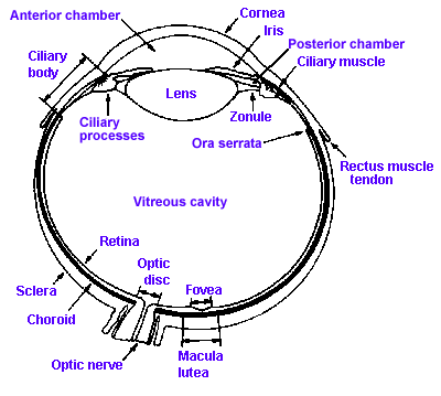 |
Fig. 7-1. A section through the human eye illustrating the major structures. (Walls GL: The Vertebrate Eye and its Adaptive Radiations. New York, Hafner, 1967) |
Figure 7-1 shows a cross section through the human eye. It consists of two fluid-filled chambers separated by a transparent structure, the lens. Nearly the entire eye is covered with a tough, fibrous coating called the sclera that is modified anteriorly to form the transparent cornea. The human cornea is about 12 mm in diameter, about 0.5 mm thick in the center and 0.75 to 1 mm thick on the edge, and it is made of the same collagenous connective tissue substance as is the sclera, but the fibers of the cornea are oriented in parallel arrays that let light pass through with minimal scatter, whereas fibers of the sclera are random and light rays are scattered when passing through. The result is that light passes easily through the cornea, but not through the sclera. Lining the inside of the posterior two-thirds of the sclera are two membranes: the choroid, a pigment layer containing the vascular supply for the eyeball as well as mechanisms for maintaining the integrity of the photoreceptors, and the retina that contains the photoreceptors and other neural elements essential to our visual process. The fine structure of the retina will be considered in detail later.
The human lens is about 11 mm in diameter and 3.5 mm thick at its thickest point, and it is suspended in place by zonule fibers that attach to the ciliary process anterior to the retina. A set of smooth muscle fibers, the ciliary muscle, lie between the ciliary process and sclera. Just anterior to the lens is a pigmented structure called the iris, that is like the diaphragm on some cameras in that it has a hole in the center of variable aperture, the pupil. The pupil is surrounded by two sets of muscles, one that encircles the aperture, the sphincter pupillae, and one that runs radially out from it, the dilator pupillae.
The anterior chamber of the eye is filled with aqueous humor, a watery fluid of low protein content that is formed from plasma. The vitreous cavity contains a gelatinous substance, the vitreous or vitreous humor. In many people the vitreous is not completely clear, but contains particulate matter that is not transparent. This material may be stationery or may float around, producing „spots before the eyes,“ the floating variety being called „floaters.“
The entire article can be found here.