Category: articles

How artificial light is wrecking your sleep, and what to do about it

2013 article

Source Chris Kresser chriskresser.com

“A good laugh and a long sleep are the best cures in the doctor’s book.” – Irish Proverb

The evidence for the health benefits of adequate, restful sleep is overwhelming. Decades of research has shown that sleeping between 7 and 9 hours per night can relieve stress, reduce the risk of many chronic diseases, improve memory and cognitive function, and may even help with weight loss. As many of us know by now, getting adequate, high-quality sleep is one of the most important, yet under-appreciated steps you can take to improve your overall health and wellbeing.


Read more at this link.

Blue-enriched white light in the workplace improves self-reported alertness, performance and sleep quality.

2088 study

Viola AU, Surrey Sleep Research Centre, Clinical Research Centre, Egerton Road, Guildford, United Kingdom
James LM, Schlangen LJ, Dijk DJ

OBJECTIVES:

Specifications and standards for lighting installations in occupational settings are based on the spectral sensitivity of the classical visual system and do not take into account the recently discovered melanopsin-based, blue-light-sensitive photoreceptive system. The authors investigated the effects of exposure to blue-enriched white light during daytime workhours in an office setting.

METHODS:

The experiment was conducted on 104 white-collar workers on two office floors. After baseline assessments under existing lighting conditions, every participant was exposed to two new lighting conditions, each lasting 4 weeks. One consisted of blue-enriched white light (17 000 K) and the other of white light (4000 K). The order was balanced between the floors. Questionnaire and rating scales were used to assess alertness, mood, sleep quality, performance, mental effort, headache and eye strain, and mood throughout the 8-week intervention.

RESULTS:

Altogether 94 participants [mean age 36.4 (SD 10.2) years] were included in the analysis. Compared with white light (4000 K), blue-enriched white light (17 000 K) improved the subjective measures of alertness (P<0.0001), positive mood (P=0.0001), performance (P<0.0001), evening fatigue (P=0.0001), irritability (P=0.004), concentration (P<0.0001), and eye discomfort (P=0.002). Daytime sleepiness was reduced (P=0.0001), and the quality of subjective nocturnal sleep (P=0.016) was improved under blue-enriched white light. When the participants‘ expectation about the effect of the light treatments was entered into the analysis as a covariate, significant effects persisted for performance, alertness, evening fatigue, irritability, difficulty focusing, concentrating, and blurred vision.

CONCLUSIONS:

Exposure to blue-enriched white light during daytime workhours improves subjective alertness, performance, and evening fatigue.


The study can be found at this link.

Marc Green Phd – Night vision

2013 article

Marc Green Phd.

Night vision is an important factor in understanding the cause of accidents that occur under low visibility. Here, I briefly outline some basics, roughly what I would expect my students to know at the end of an introductory perception course. [See related articles The Invisible Pedestrian and Police Shootings.]

Photopic, Mesopic and Scotopic Vision

Humans can see over a light intensity range of several million to one. In order to achieve this extraordinary feat while maintaining good contrast sensitivity, the eye adjusts to the prevailing conditions and changes its mode of operation as light levels decline from day to night.Every beginner’s textbook discusses rods and cones, so amateurs pick up on this these terms and focus too heavily on them. Photoreceptors alone are insufficient to explain night vision. Moreover, rod vision and night vision are not synonymous. The more important concept is „receptive field,“ which is fundamental to all visual processing. Anyone who claims to be an expert in vision/perception must have a thorough understanding of receptive fields, their various types, how they operate, how they change with conditions and how they determine visual capability. I won’t go into a full explanation of receptive fields because it is too large a topic. However, I will mention two of their properties, inhibition and convergence.

Individual cones and rods have very similar sensitivity to light. Both respond to a single quantum of light, although rods produce a bigger response. A major difference between day and night vision is inhibition and convergence, the way the photoreceptors are wired together, and the amount of light-sensitive photopigment available. Moreover, most „night vision“ occurs in a mixed rod/cone mode. The overall operation of the eye in diminishing light levels is better described in terms of three operating modes, photopic, mesopic and scotopic. Photopic vision occurs at high light levels and is characterized by 1) cone photoreceptors, 2) low light sensitivity, 3) high acuity and 4) color vision. Scotopic vision occurs at very low light levels and exhibits 1) use of rod photoreceptors, 2) high light sensitivity, 3) poor acuity and 4) no color vision.
The entire article can be found at this link.

Bright Lights, Big Problems – Sky & telescope article

2006 article

J. Kelly Beatty and Rachel Thessin

Like death and taxes, there’s no denying that outdoor lighting has become an inescapable part of life. Streetlights adorn our roads, billboards stud our freeways, shopping-center parking lots are aglow from dusk to dawn, businesses obsess over late-night security, and convenience stores outdazzle one another to compete for customers. We cheat the night of darkness and, in the process, create light pollution that robs the sky of stars.

Electric streetlights have been with us since the 1880s, and it wasn’t long thereafter that some manufacturers recognized the visual and cost-saving benefits of directing light down, onto the ground. In 1918 the Holophane Glass Co. published the very first roadway-lighting manual. Titled The New Era in Street Lighting, it set forth a number of recommended practices, among them the common-sense notion that “Light above the horizontal must be conserved.” In a later section, the manual notes:

In addition to the two fundamental items of highly efficient lamps and the effective use of the light, as discussed, it is very important to see to it that the street lighting system produces an effect which surrounds the eyes of those using the streets with conditions under which the eye is free to perform its functions properly. Any system which fails in this respect is extravagant — no matter how efficient the lamps nor how efficiently the light may be directed upon the street surfaces or objects. Glare serves seriously to reduce the discerning power of the eye.

Unfortunately, almost no one heeded this unsung champion of good lighting practices. Instead, artificial skyglow became markedly more obvious in the late 20th century with the widespread use of high-intensity fixtures utilizing mercury-vapor and high-pressure-sodium lamps, and with a societal shift that found more people on the streets at night — and at later hours — than ever before. As our nocturnal wanderings increased, so too did the need for ubiquitous nighttime illumination. Then decision-makers began to equate “more light” with “better safety and security,” even though objective proof of such a relationship did not exist.
The entire article can be found at this link.

American Medical Association Addresses Light Pollution

2012 article

Camille M. Carlisle

Researchers are raising several possible health concerns related to nighttime light exposure, among them a higher risk of cancer.

I usually think of light pollution as astronomers’ concern. Who else would mind if the sky glow is so bright that it washes out Orion? (When I can’t see Orion, I feel jilted — yes, even in the months when it’s below the horizon at night.) But the issue has a broader reach than my petulance. Fighting light pollution isn’t merely about seeing stars; it’s about being sensible in our usage and reducing waste.
The document’s first human concern is glare, which report coauthor Dr. Mario Motta (Tufts Medical School) outlined for S&T readers back in 2009. Glare’s a pretty standard discussion topic in light-pollution conversations, in part because, as drivers age, their eyes become less able to cope with poorly directed light that scatters inside the eye itself. In 2009 the AMA passed a resolution submitted by Motta supporting the use of fully shielded lights, such as the flat-bottomed street lights. The new report reaffirms that resolution.Still, I was surprised to see that the American Medical Association recently released a report entitled “Light Pollution: Adverse Health Effects of Nighttime Lighting.” It’s a review of some of the available research literature on nighttime lighting’s effect on people; it doesn’t present new research done by the AMA, although many of the results considered come from the authors‘ own work. The report covers a lot of ground, but it’s unclear what the review’s effect will ultimately be.

Vision researcher Gary Rubin (University College London) agrees with the report’s concern, saying the conclusions are “balanced, well-reasoned and thoroughly researched.” Disability glare — as opposed to “discomfort glare,” which differs from person to person — is definitely a problem for drivers, he says, noting that some cataract patients have had second surgeries to replace their new intraocular lenses with another kind that causes less nighttime glare. And as many of us know from experience, modern halogen and LED headlamps can make nighttime driving downright painful. (I can’t tell you how many times I’ve looked away from an oncoming car’s bright bluish headlights and thought, with scathing condescension, “Is that really necessary?”) Blue-rich light’s destructive effect on the molecule rhodopsin (a.k.a. “visual purple”) in the retina is what makes these headlights hurt so much.
The entire article can be found at this link.

The Color-Sensitive Cones

The Color-Sensitive Cones

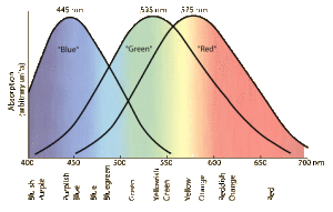
In 1965 came experimental confirmation of a long expected result – there are three types of color-sensitive cones in the retina of the human eye, corresponding roughly to red, green, and blue sensitive detectors.

colcon

More information here.

Vision

2011 article

Source michaeldmann.net

Photoreception is a particularly important sense for most primates, including man, but it is not unique to primates or even mammals. Even mollusks have photoreceptors, but one may question whether they possess vision in the same sense as we have it. Most objects reflect light, and because light travels at high speed, it is possible to nearly instantly assess their shape, size, position, speed, and direction of movement. The light rays emanating from an object are gathered and focused onto an array of photoreceptors. Activities generated in the different photoreceptors by the light interact to produce a two-dimensional representation of the object which is transmitted to the brain. The brain then reconstructs a three-dimensional representation using information received from the two eyes. The end-products of the activity of the visual system are sensations that represent the object and its surroundings. These sensations can be used to guide our immediate behavior, or they can be stored for future reference. Visual sensations contain a great deal of information, and understanding these complex phenomena is no simple matter. The best place to begin the study of vision is at the eye itself.

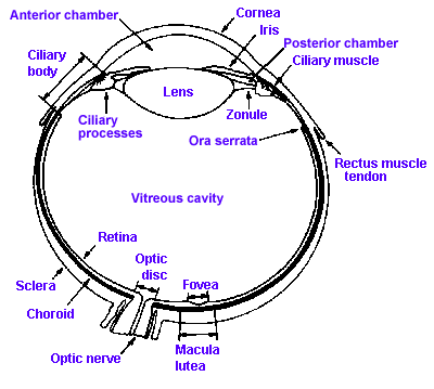
Fig. 7-1. A section through the human eye illustrating the major structures. (Walls GL: The Vertebrate Eye and its Adaptive Radiations. New York, Hafner, 1967)

Figure 7-1 shows a cross section through the human eye. It consists of two fluid-filled chambers separated by a transparent structure, the lens. Nearly the entire eye is covered with a tough, fibrous coating called the sclera that is modified anteriorly to form the transparent cornea. The human cornea is about 12 mm in diameter, about 0.5 mm thick in the center and 0.75 to 1 mm thick on the edge, and it is made of the same collagenous connective tissue substance as is the sclera, but the fibers of the cornea are oriented in parallel arrays that let light pass through with minimal scatter, whereas fibers of the sclera are random and light rays are scattered when passing through. The result is that light passes easily through the cornea, but not through the sclera. Lining the inside of the posterior two-thirds of the sclera are two membranes: the choroid, a pigment layer containing the vascular supply for the eyeball as well as mechanisms for maintaining the integrity of the photoreceptors, and the retina that contains the photoreceptors and other neural elements essential to our visual process. The fine structure of the retina will be considered in detail later.

The human lens is about 11 mm in diameter and 3.5 mm thick at its thickest point, and it is suspended in place by zonule fibers that attach to the ciliary process anterior to the retina. A set of smooth muscle fibers, the ciliary muscle, lie between the ciliary process and sclera. Just anterior to the lens is a pigmented structure called the iris, that is like the diaphragm on some cameras in that it has a hole in the center of variable aperture, the pupil. The pupil is surrounded by two sets of muscles, one that encircles the aperture, the sphincter pupillae, and one that runs radially out from it, the dilator pupillae.

The anterior chamber of the eye is filled with aqueous humor, a watery fluid of low protein content that is formed from plasma. The vitreous cavity contains a gelatinous substance, the vitreous or vitreous humor. In many people the vitreous is not completely clear, but contains particulate matter that is not transparent. This material may be stationery or may float around, producing „spots before the eyes,“ the floating variety being called „floaters.“
The entire article can be found here.

Opponent melanopsin and S-cone signals in the human pupillary light response

2014 study

Manuel Spitschan, Sandeep Jain, David H. Brainard, and Geoffrey K. Aguirre. Departments of Psychology and Neurology, University of Pennsylvania, Philadelphia, PA 19104

In the human, cone photoreceptors (L, M, and S) and the melanopsincontaining, intrinsically photosensitive retinal ganglion cells (ipRGCs) are active at daytime light intensities. Signals from cones are combined both additively and in opposition to create the perception of overall light and color. Similar mechanisms seem to be at work in the control of the pupil’s response to light. Uncharacterized however, is the relative contribution of melanopsin and S cones, with their overlapping, short-wavelength spectral sensitivities. We measured the response of the human pupil to the separate stimulation of the cones and melanopsin at a range of temporal frequencies under photopic conditions. The S-cone and melanopsin photoreceptor channels were found to be low-pass, in contrast to a band-pass response of the pupil to L- and M-cone signals. An examination of the phase relationships of the evoked responses revealed that melanopsin signals add with signals from L and M cones but are opposed by signals from S cones in control of the pupil. The opposition of the S cones is revealed in a seemingly paradoxical dilation of the pupil to greater S-cone photon capture. This surprising result is explained by the neurophysiological properties of ipRGCs found in animal studies.

Results Using an infrared camera, we measured the consensual PLR of human participants while they observed sinusoidal modulations in the spectrum of a light (Fig. 1B). The stimulus modulations were designed to target specific photoreceptors. The cones and melanopsin have different but overlapping spectral sensitivities. Despite the overlap, it is possible to create sets of light spectra such that the absorption of photons is constant for all of the photoreceptor classes except one (14–16) (Fig. 1C). Modulation between a pair of these “silent substitution” spectra increases and decreases the response of (for example) melanopsin-containing ipRGCs while maintaining nominally constant stimulation of the cones. Separate modulations were designed for melanopsin, S cones, and L+M cones together (a modulation that varied luminance as well as chromaticity). An isochromatic modulation (melanopsin+S+M+L) was also used. All modulations were designed to produce 50% contrast on their targeted photoreceptor(s). Rods were silenced by modulating the spectra about a photopic background (∼800 cd/m2 ). The stimulus was wide-field (27.5°), spatially uniform, and had the central 5° obscured to avoid variation in photoreceptor spectral sensitivity across the visual field caused by the presence of the foveal macular pigment (17). Simulations and control experiments support the specificity of the photoreceptor isolation (Figs. S1–S5 and Table S1). We measured pupil responses from 16 subjects while they observed the different photoreceptor-directed modulations at two for each combination of photoreceptor target and modulation frequency. The two-filter model fits the average amplitude and phase data (Fig. 5A) with parameters similar to those found for subject 01 (Table S2). When expressed as a polar plot (Fig. 5B), the agreement between the group data and model fits is apparent. Interestingly, there is systematic “rotation” of the phase of both the pupil brightness and S-cone responses at the lower temporal frequency that is not captured by the model. This may result from individual differences in the phase of S-cone responses at low temporal frequencies, as is seen between subject 01 and subject 02 (Fig. 4), because the average data do not fully constrain the model and the fits shown are based on parameters obtained for subject 01.

Whole study on this link

Melanopsin and Rod-Cone Photoreceptors Play Different Roles in Mediating Pupillary Light Responses during Exposure to Continuous Light in Humans

2012 article

Joshua J. Gooley,1,2,3 Ivan Ho Mien,4 Melissa A. St. Hilaire,2,3 Sing-Chen Yeo,5 Eric Chern-Pin Chua,1 Eliza van Reen,2,3 Catherine J. Hanley,2 Joseph T. Hull,2,3 Charles A. Czeisler,2,3 and Steven W. Lockley2,3

1 Program in Neuroscience and Behavioral Disorders, Duke–National University of Singapore Graduate Medical School Singapore, Singapore 169857,

2 Division of Sleep Medicine, Department of Medicine, Brigham and Women’s Hospital, and

3 Division of Sleep Medicine, Department of Medicine, Harvard Medical School, Boston, Massachusetts 02115, 4 Graduate School for Integrative Sciences and Engineering, National University of Singapore, Singapore 117456, and 5 National Neuroscience Institute, Singapore 308433

In mammals, the pupillary light reflex is mediated by intrinsically photosensitive melanopsin-containing retinal ganglion cells that also receive input from rod– cone photoreceptors. To assess the relative contribution of melanopsin and rod– cone photoreceptors to the pupillary light reflex in humans, we compared pupillary light responses in normally sighted individuals (n  24) with a blind individual lacking rod– cone function. Here, we show that visual photoreceptors are required for normal pupillary responses to continuous light exposure at low irradiance levels, and for sustained pupillary constriction during exposure to light in the long-wavelength portion of the visual spectrum. Inthe absence of rod– conefunction, pupillomotor responses are slow and sustained, and cannottrackintermittent light stimuli, suggesting that rods/cones are required for encoding fast modulations in light intensity. In sighted individuals, pupillary constriction decreased monotonically for at least 30 min during exposureto continuous low-irradiance light, indicatingthat steady-state pupillary responses are an order of magnitude slower than previously reported. Exposure to low-irradiance intermittent green light (543 nm; 0.1– 4 Hz)for 30 min, which was givento activate cone photoreceptors repeatedly, elicited sustained pupillary constriction responses that were more than twice as great compared with exposure to continuous green light. Our findings demonstrate nonredundant roles for rod– cone photoreceptors and melanopsin in mediating pupillary responses to continuous light. Moreover, our results suggest that it might be possible to enhance nonvisual light responses to low-irradiance exposures by using intermittent light to activate cone photoreceptors repeatedly in humans.

Introduction

The pupillary light reflex (PLR) regulates the amount of light that reaches the retina. In doing so, the PLR optimizes visual acuity over a wide range of illuminance levels (Campbell and Gregory, 1960) and protects the retina from the potentially damaging effects of exposure to bright light. The PLR is mediated by melanopsin-containing retinal ganglion cells that project directly to the olivary pretectal nucleus (Hattar et al., 2002; Gooley et al., 2003). Melanopsin cells are intrinsically photosensitive and respond most strongly to short-wavelength light in the blue portion of the visual spectrum (Berson et al., 2002; Dacey et al., 2005). Rod– cone photoreceptors also provide input to melanopsin cells (Belenky et al., 2003; Wong et al., 2007), but melanopsin cells are not required for pattern-forming vision (Gu¨ler et al., 2008). In contrast, the PLR and other nonvisual light responses are abolished if rod– cone and melanopsin signaling pathways are disrupted simultaneously (Hattar et al., 2003; Panda et al., 2003), or if melanopsin cells are selectively killed (Gu¨ler et al., 2008). In humans, rods and cones are capable of driving the initial rapid constriction of the pupils in response to light (Alpern and Campbell, 1962), whereas the PLR is most sensitive to shortwavelength blue light during exposure to continuous light (Bouma, 1962; Alexandridis and Koeppe, 1969; Mure et al., 2009; McDougal and Gamlin, 2010), even in the absence of rod and cone function (Zaidi et al., 2007), suggesting a primary role for melanopsin photopigment. For light intensities below the threshold of activation for melanopsin cells, spectral responses of the PLR during exposure to continuous light are consistent with a role for rods, with little or no contribution from cones (McDougal and Gamlin, 2010). By comparison, attempts to isolate visual photoreceptor contributions to the PLR using the method of silent substitution have yielded contrasting results, with one study reporting a contribution from M- and L-cones (Tsujimura et al., 2010), and another reporting a possible contribution from rod photoreceptors (Vie´not et al., 2010). Melanopsin and rod–cone contributions to the PLR are difficult to assess in normally sighted humans due to overlap of spectral sensitivity for the various photoreceptor types. As demonstrated in macaques, which have trichromatic vision similar to humans, melanopsin-dependent pupillary responses can be examined in isolation when rod– cone signaling is disrupted (Gamlin et al., 2007). Hence, the role of melanopsin versus rod– cone photoreceptors in driving pupillary light responses could potentially be assessed in blind humans with complete loss of visual function, but with preservation of the retinal ganglion cell layer and melanopsin function (Czeisler et al., 1995; Klerman et al., 2002; Zaidi et al., 2007). To date, however, fewer than a dozen such patients have been identified worldwide. In the present study, we provide a detailed analysis of pupillary light responses in a patient with intact nonvisual responses to light (Zaidi et al., 2007), but without a functional outer retina. Here, the relative contribution of melanopsin and visual photoreceptors was assessed by comparing PLR responses in the totally visually blind patient with normally sighted individuals.

Full article here

Exposure to Room Light before Bedtime Suppresses Melatonin Onset and Shorens Melatonin Duration in Humans

2010 article

Joshua J. Gooley, Kyle Chamberlaine, Kurt A. Smith, Sat Bir S. Khalsa, Shantha M. W. Rajaratnam, Eliza Van Reen, Jamie M. Zeitzer, Charles A. Czeisler, and Steven W. Lockley

Abstract

Context:

Millions of individuals habitually expose themselves to room light in the hours before bedtime, yet the effects of this behavior on melatonin signaling are not well recognized.

Objective:

We tested the hypothesis that exposure to room light in the late evening suppresses the onset of melatonin synthesis and shortens the duration of melatonin production.

Design:

In a retrospective analysis, we compared daily melatonin profiles in individuals living in room light (<200 lux) vs. dim light (<3 lux).

Patients:

Healthy volunteers (n = 116, 18–30 yr) were recruited from the general population to participate in one of two studies.

Setting:

Participants lived in a General Clinical Research Center for at least five consecutive days.

Intervention:

Individuals were exposed to room light or dim light in the 8 h preceding bedtime.

Outcome Measures:

Melatonin duration, onset and offset, suppression, and phase angle of entrainment were determined.

Results:

Compared with dim light, exposure to room light before bedtime suppressed melatonin, resulting in a later melatonin onset in 99.0% of individuals and shortening melatonin duration by about 90 min. Also, exposure to room light during the usual hours of sleep suppressed melatonin by greater than 50% in most (85%) trials.

Conclusions:

These findings indicate that room light exerts a profound suppressive effect on melatonin levels and shortens the body’s internal representation of night duration. Hence, chronically exposing oneself to electrical lighting in the late evening disrupts melatonin signaling and could therefore potentially impact sleep, thermoregulation, blood pressure, and glucose homeostasis.

full article here