Category: articles

Pupillary light reflex

The pupillary light reflex (PLR) is a reflex that controls the diameter of the pupil, in response to the intensity (luminance) of light that falls on the retina of the pupil and the eye, thereby assisting in adaptation to various levels of lightness/darkness. A greater intensity of light causes the pupil to constrict (miosis/myosis) (allowing less light in), whereas a lower intensity of light causes the pupil to dilate (mydriasis,expansion) (allowing more light in). Thus, the pupillary light reflex regulates the intensity of light entering the eye.

The pupillary light reflex pathway has an afferent limb (CN II) and efferent limb (CN III). The ganglion cells of the retina project bilaterally to the pretectal nuclei. The pretectal nuclei projects crossed and uncrossed fibers to the Edinger-Westphal nucleus, which gives rise to the preganglionic parasympathetic fibers. These fibers exit the midbrain with CN III and synapse with postganglionic parasympathetic neurons of the ciliary ganglion, which innervates the sphincter muscle of the iris.

more on this link.

Caveman: An Interview with Michel Siffre

Article from 2008

Michel Siffre - scientist based in Paris, author of several books, including Beyond Time (McGraw-Hill, 1964) and Découvertes dans les grottes mayas (Arthaud, 1993).

Joshua Foer - freelance science writer.

In 1962, a French speleologist named Michel Siffre spent two months living in total isolation in a
subterranean cave, without access to clock, calendar, or sun. Sleeping and eating only when his body told
him to, his goal was to discover how the natural rhythms of human life would be affected by living
“beyond time.” Over the next decade, Siffre organized over a dozen other underground time isolation
experiments, before he himself returned to a cave in Texas in 1972 for a six-month spell. His work helped
found the field of human chronobiology. Joshua Foer interviewed Siffre by email.

Whole interview here.

A neural mechanism for exacerbation of headache by light

2010 study

Rodrigo Noseda,1 Vanessa Kainz,1 Moshe Jakubowski,1Joshua J. Gooley,2 Clifford B. Saper,2,3Kathleen Digre,4 and Rami Burstein1,3

1Department of Anesthesia, Harvard Medical School, Boston, Massachusetts, USA
2Department of Neurology, Harvard Medical School, Boston, Massachusetts, USA
3Beth Israel Deaconess Medical Center and Program in Neuroscience, Harvard Medical School, Boston, Massachusetts, USA
4Department of Neurology and Ophthalmology, Moran Eye Center, University of Utah, Salt Lake City, Utah

The perception of migraine headache, which is mediated by nociceptive signals transmitted from the cranial dura mater to the brain, is uniquely exacerbated by exposure to light. Here we show that exacerbation of migraine headache by light is prevalent among blind persons who maintain non-image-forming photoregulation in the face of massive rod/cone degeneration. Using single-unit recording and neural tract-tracing in the rat, we identified dura-sensitive neurons in the posterior thalamus, whose activity was distinctly modulated by light, and whose axons projected extensively across layers I through V of somatosensory, visual and associative cortices. The cell bodies and dendrites of such dura/light-sensitive neurons were apposed by axons originating from retinal ganglion cells, predominantly from intrinsically-photosensitive retinal ganglion cells – the principle conduit of non-image-forming photoregulation. We propose that photoregulation of migraine headache is exerted by a non-image-forming retinal pathway that modulates the activity of dura-sensitive thalamocortical neurons.

Whole study on this link.

Human pineal physiology and functional significance of melatonin

2004 study

M. Mila Macchia, Jeffrey N. Bruceb

a New York State Psychiatric Institute, College of Physicians and Surgeons, Columbia University, 1051 Riverside Drive, Unit 50, New York, NY 10032, United States
b Bartoli Brain Tumor Research Laboratory, Neurological Institute, Columbia Presbyterian Medical Center New York, NY, United States

Abstract

Descriptions of the pineal gland date back to antiquity, but its functions in humans are still poorly understood. In both diurnal and nocturnal vertebrates, its main product, the hormone melatonin, is synthesized and released in rhythmic fashion, during the dark portion of the day–night cycle. Melatonin production is controlled by an endogenous circadian timing system and is also suppressed by light. In lower vertebrates, the pineal gland is photosensitive, and is the site of a self-sustaining circadian clock. In mammals, including humans, the gland has lost direct photosensitivity, but responds to light via a multisynaptic pathway that includes a subset of retinal ganglion cells containing the newly discovered photopigment, melanopsin. The mammalian pineal also shows circadian oscillations, but these damp out within a few days in the absence of input from the primary circadian pacemaker in the suprachiasmatic nuclei (SCN). The duration of the nocturnal melatonin secretory episode increases with nighttime duration, thereby providing an internal calendar that regulates seasonal cycles in reproduction and other functions in photoperiodic species. Although humans are not considered photoperiodic, the occurrence of seasonal affective disorder (SAD) and its successful treatment with light suggest that they have retained some photoperiodic responsiveness. In humans, exogenous melatonin has a soporific effect, but only when administered during the day or early evening, when endogenous levels are low. Some types of primary insomnia have been attributed to diminished melatonin production, particularly in the elderly, but evidence of a causal link is still inconclusive. Melatonin administration also has mild hypothermic and hypotensive effects. A role for the pineal in human reproduction was initially hypothesized on the basis of clinical observations on the effects of pineal tumors on sexual development. More recent data showing an association between endogenous melatonin levels and the onset of puberty, as well as observations of elevated melatonin levels in both men and women with hypogonadism and/or infertility are consistent with such a hypothesis, but a regulatory role of melatonin has yet to be established conclusively. A rapidly expanding literature attests to the involvement of melatonin in immune function, with high levels promoting and low levels suppressing a number of immune system parameters. The detection of melatonin receptors in various lymphoid organs and in lymphocytes suggests multiple mechanisms of action. Melatonin has been shown to be a powerful antioxidant, and has oncostatic properties as well, both direct and indirect, the latter mediated by its effects on reproductive hormones. Finally, there are reports of abnormal daily melatonin profiles in a number of psychiatric and neurological disorders, but the significance of such abnormalities is far from clear.

Whole study here

Sleep in the Critically ill Patient

2006 study

Gerald L. Weinhouse, MD1 ; Richard J. Schwab, MD2

1 The Division of Pulmonary and Critical Care Medicine, Brigham and Women’s Hospital, Boston, MA;

2 the Division of Pulmonary, Critical Care, and Sleep Medicine, University of Pennsylvania Medical Center, Philadelphia, PA

Abstract:

Critically ill patients are known to suffer from severely fragmented sleep with a predominance of stage I sleep and a paucity of slow wave and REM sleep. The causes of this sleep disruption include the intensive care unit (ICU) environment, medical illness, psychological stress, and many of the medications and other treatments used to help those who are critically ill. The clinical importance of this type of sleep disruption in critically ill patients, however, is not known. This article reviews the literature on sleep disruption in the ICU, the effects of sepsis on sleep, the effects of commonly used ICU medications on sleep, the relationship between sleep and sedation, and the literature on the biological and psychological consequences of sleep deprivation specifically as it relates to the critically ill. Finally, an integrative approach to improving sleep in the ICU is described.

Whole study here

Melanopsin Ganglion Cells: A Bit of Fly in the Mammalian Eye by Dustin M. Graham

2011 article

Dustin M. Graham

Dr. Dustin M. Graham was born and raised in Pleasanton, California and received his Ph.D. in neuroscience from Brown University. He began his research career at Santa Clara University in the lab of Dr. David Tauck, studying neural pathways of learning and memory in the pond snail Lymnaea stagnalis. His interests turned to the retina while working with Dr. Ralph Nelson at the National Institutes of Health. There he helped develop a rapid labeling technique to delineate morphological subtypes of retinal neurons in zebrafish. As a graduate student, Dustin studied mammalian circadian rhythms and the newly discovered melanopsin ganglion cell activity in Dr. David Berson’s lab. He focussed on the phototransduction cascade in ipRGCs, and developed a dissociation and culturing procedure to identify and record light responses from isolated ipRGCs. Dustin is currently a post-doctoral research fellow in the Psychology department at the University of Virginia where he studies development and synaptic mechanisms of the gustatory system in rats.

Introduction.

For the greater part of 150 years it was assumed that the mammalian retina contained only two types of photoreceptors; rods and cones. However, a flurry of recent evidence has demonstrated the existence of a third type of mammalian photoreceptor that differs greatly from rods and cones. This type utilizes a different photopigment, is much less sensitive to light, and has far less spatial resolution; characteristics that fit perfectly with this photoreceptor’s primary function of signaling changes in ambient light levels to the brain throughout the day. Most surprisingly, these photoreceptors are ganglion cells, and thus, have the unique ability to communicate directly with the brain. These intrinsically photosensitive retinal ganglion cells (ipRGCs) are a rare sub-population of ganglion cells (1-3%) whose primary role is to signal light for unconscious visual reflexes, such as pupillary constriction, and regulating a number of daily behavioral and physiological rhythms, collectively called circadian rhythms. This latter process, which adjusts circadian rhythms to the light/dark cycle of an animal’s environment, is known as photoentrainment. The visual behaviors under ipRGC control are remarkably tonic, and require long integration times of ambient light levels. The unique properties of ipRGCs, both functionally and anatomically, make them well suited for regulating such behaviors.

Whole article here

Opponent melanopsin and S-cone signals in the human pupillary light response

2014 study

Manuel Spitschana , Sandeep Jainb , David H. Brainarda,1, and Geoffrey K. Aguirreb,1

Departments of a Psychology and b Neurology, University of Pennsylvania, Philadelphia, PA 19104

Edited by Dennis M. Dacey, The University of Washington, Seattle, WA, and accepted by the Editorial Board September 12, 2014 (received for review January 17, 2014)

In the human, cone photoreceptors (L, M, and S) and the melanopsincontaining, intrinsically photosensitive retinal ganglion cells (ipRGCs) are active at daytime light intensities. Signals from cones are combined both additively and in opposition to create the perception of overall light and color. Similar mechanisms seem to be at work in the control of the pupil’s response to light. Uncharacterized however, is the relative contribution of melanopsin and S cones, with their overlapping, short-wavelength spectral sensitivities. We measured the response of the human pupil to the separate stimulation of the cones and melanopsin at a range of temporal frequencies under photopic conditions. The S-cone and melanopsin photoreceptor channels were found to be low-pass, in contrast to a band-pass response of the pupil to L- and M-cone signals. An examination of the phase relationships of the evoked responses revealed that melanopsin signals add with signals from L and M cones but are opposed by signals from S cones in control of the pupil. The opposition of the S cones is revealed in a seemingly paradoxical dilation of the pupil to greater S-cone photon capture. This surprising result is explained by the neurophysiological properties of ipRGCs found in animal studies.

Whole study on this link

Melanopsin and Rod–Cone Photoreceptors Play Different Roles in Mediating Pupillary Light Responses during Exposure to Continuous Light in Humans

2012 study

Joshua J. Gooley,1,2,3 Ivan Ho Mien,4 Melissa A. St. Hilaire,2,3 Sing-Chen Yeo,5 Eric Chern-Pin Chua,1 Eliza van Reen,2,3 Catherine J. Hanley,2 Joseph T. Hull,2,3 Charles A. Czeisler,2,3 and Steven W. Lockley2,3

1 Program in Neuroscience and Behavioral Disorders, Duke–National University of Singapore Graduate Medical School Singapore, Singapore 169857,
2 Division of Sleep Medicine, Department of Medicine, Brigham and Women’s Hospital, and 3 Division of Sleep Medicine, Department of Medicine, Harvard Medical School, Boston, Massachusetts 02115, 4 Graduate School for Integrative Sciences and Engineering, National University of Singapore, Singapore 117456, and 5 National Neuroscience Institute, Singapore 308433

In mammals, the pupillary light reflex is mediated by intrinsically photosensitive melanopsin-containing retinal ganglion cells that also
receive input from rod– cone photoreceptors. To assess the relative contribution of melanopsin and rod– cone photoreceptors to the
pupillary light reflex in humans, we compared pupillary light responses in normally sighted individuals (n  24) with a blind individual
lacking rod– cone function. Here, we show that visual photoreceptors are required for normal pupillary responses to continuous light
exposure at low irradiance levels, and for sustained pupillary constriction during exposure to light in the long-wavelength portion of the
visual spectrum. Inthe absence of rod– conefunction, pupillomotor responses are slow and sustained, and cannottrackintermittent light
stimuli, suggesting that rods/cones are required for encoding fast modulations in light intensity. In sighted individuals, pupillary
constriction decreased monotonically for at least 30 min during exposureto continuous low-irradiance light, indicatingthat steady-state
pupillary responses are an order of magnitude slower than previously reported. Exposure to low-irradiance intermittent green light (543
nm; 0.1– 4 Hz)for 30 min, which was givento activate cone photoreceptors repeatedly, elicited sustained pupillary constriction responses
that were more than twice as great compared with exposure to continuous green light. Our findings demonstrate nonredundant roles for
rod– cone photoreceptors and melanopsin in mediating pupillary responses to continuous light. Moreover, our results suggest that it
might be possible to enhance nonvisual light responses to low-irradiance exposures by using intermittent light to activate cone photoreceptors
repeatedly in humans.

Whole study here

Circadian Rhythms Fact Sheet

2012 article

National Institute of General Medical Science

NIGMS is a part of the National Institutes of Health that supports basic research to increase our understanding of biological processes and lay the foundation for advances in disease diagnosis, treatment and prevention.

What are circadian rhythms?

Circadian rhythms are physical, mental and behavioral changes that follow a roughly 24-hour cycle, responding primarily to light and darkness in an organism’s environment. They are found in most living things, including animals, plants and many tiny microbes. The study of circadian rhythms is called chronobiology.

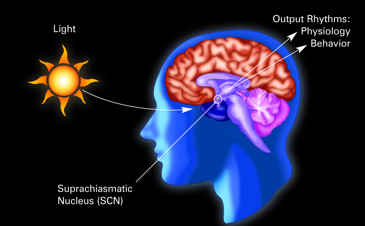
Illustration of a human brain with the suprachiasmatic nucleus labelled.

Are circadian rhythms the same thing as biological clocks?

No, but they are related. Our biological clocks drive our circadian rhythms.

What are biological clocks?

The biological clocks that control circadian rhythms are groupings of interacting molecules in cells throughout the body. A „master clock“ in the brain coordinates all the body clocks so that they are in synch.

What is the master clock?

NATIONAL INSTITUTES OF HEALTH SLEEP DISORDERS RESEARCH PLAN

2011 study

National Center On Sleep Disorders Research

Sleep and circadian disturbances and disorders affect millions of Americans across all demographic groups. An estimated 25-30% of the general adult population, and a comparable percentage of children and adolescents, is affected by decrements in sleep health that are proven contributors to disability, morbidity, and mortality. As a result, sleep and circadian disturbances and disorders have been recognized by Congress and the Department of Health and Human Services1,2 as high priority targets for basic and clinical scientific investigation. Three general categories of sleep and circadian disorders and disturbances have been described: 1) disorders of sleep and circadian rhythms; 2) sleep deficiency; and 3) environmental disruption of circadian functions. In addition to clinical sleep and circadian disorders, sleep deficiency and circadian disruption resulting from lifestyle factors are increasingly common societal problems that increase disease risk through complex pathways. Advances sweeping across the spectrum of biomedical inquiry have transformed the sleep and circadian research landscape since the first NIH Sleep Disorders Research Plan was developed in 1996. The scientific domain is well-poised today to contribute knowledge advances and emerging technologies to the goals of understanding mechanisms of disease risk, accelerating translation from bench to bedside to community, and developing the evidence based evaluation of intervention effectiveness. Opportunities for research training exist in all areas of sleep and circadian biology and at multiple levels of the educational ladder. Scientific cross-fertilization and the development of an interdisciplinary workforce would stimulate the application of sleep and circadian scientific advances in cross-cutting domains.
Whole study here